ESI vs MALDI: A Decision Guide for Peptide Analysis
Discover when to choose ESI or MALDI for peptide analysis. Learn the advantages and best applications of each method to maximize your results.
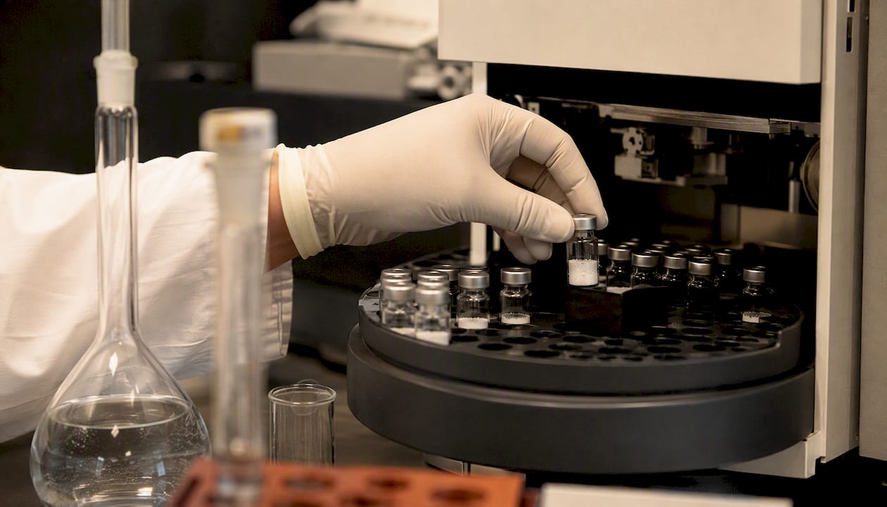
For LC-based peptide sequencing and quantitative workflows, choose LC-ESI-MS. For rapid profiling, high-throughput plate analysis, and spatial imaging, choose MALDI or MALDI-TOF. That single rule resolves most method-selection questions that cross an analytical scientist’s bench, but the exceptions matter enough to spell out before you commit instrument time.
Run MALDI when your sample carries heavy salt contamination you can’t easily clean up, when you need a fast intact-mass check on many spots, or when spatial distribution in tissue is the actual research question. Run ESI, coupled to liquid chromatography, when you need multiply charged precursors for MS/MS-rich sequencing, when quantitation has to hold up across a validated pipeline, or when your analyte mixture is complex.
- Pick ESI when: you need online LC separation, multiply charged precursors for fragmentation, or defensible quantitation.
- Pick MALDI when: you need speed, salt tolerance, plate-based throughput, or spatial imaging.
- Run both when: proteome coverage matters more than turnaround time, since the two methods disagree on which peptides they detect more often than most scientists expect.
Statistic: A large-scale proteomic study examining more than 72,000 peptides found that only about 39% were identified by both MALDI and ESI. The two techniques aren’t redundant checks on each other. They’re catching different halves of the same sample.
Key Takeaways
The choice between ESI and MALDI comes down to whether your experiment needs online separation and MS/MS depth or offline speed and salt tolerance, and roughly 39% peptide-ID overlap means the two methods often see different halves of the same sample.
| Point | Details |
|---|---|
| Default to LC-ESI-MS for sequencing | Choose it when you need multiply charged precursors, MS/MS depth, or online LC separation. |
| Default to MALDI for throughput and imaging | Choose it for high-throughput plate screening, salt-tolerant samples, or spatial tissue mapping. |
| Run both for maximum coverage | Combine methods when proteome depth matters more than turnaround time, given the low peptide-ID overlap between them. |
| Match analyzer to source | Pair ESI with QTOF or Orbitrap for online workflows; pair MALDI with TOF for pulsed-ion detection. |
| Fix reproducibility at the source | Desalt samples before ESI runs and standardize matrix deposition before MALDI runs to cut variability. |
Table of Contents
- What Is the Difference Between ESI and MALDI?
- How Do ESI and MALDI Compare Across Key Performance Metrics?
- Which Ion Source Performs Best for Your Analyte Class?
- How Does the Ion Source Interact With Your Mass Analyzer?
- What Should You Expect From Quantitation and Reproducibility?
- How Do You Choose Between ESI and MALDI for Your Experiment?
- When Should You Run Both MALDI and ESI on the Same Sample?
- How Does Ionization Method Choice Fit Into Peptide QC Workflows?
- Recommended Reading on ESI vs MALDI Ionization
- Sources
What Is the Difference Between ESI and MALDI?
ESI (electrospray ionization) generates ions from a liquid stream by applying a high voltage to a flowing solvent, producing a fine spray of charged droplets that shed solvent until bare, often multiply charged, analyte ions remain. It’s built to interface directly with liquid chromatography, which is why LC-ESI-MS has become the workhorse combination for peptide and protein work.
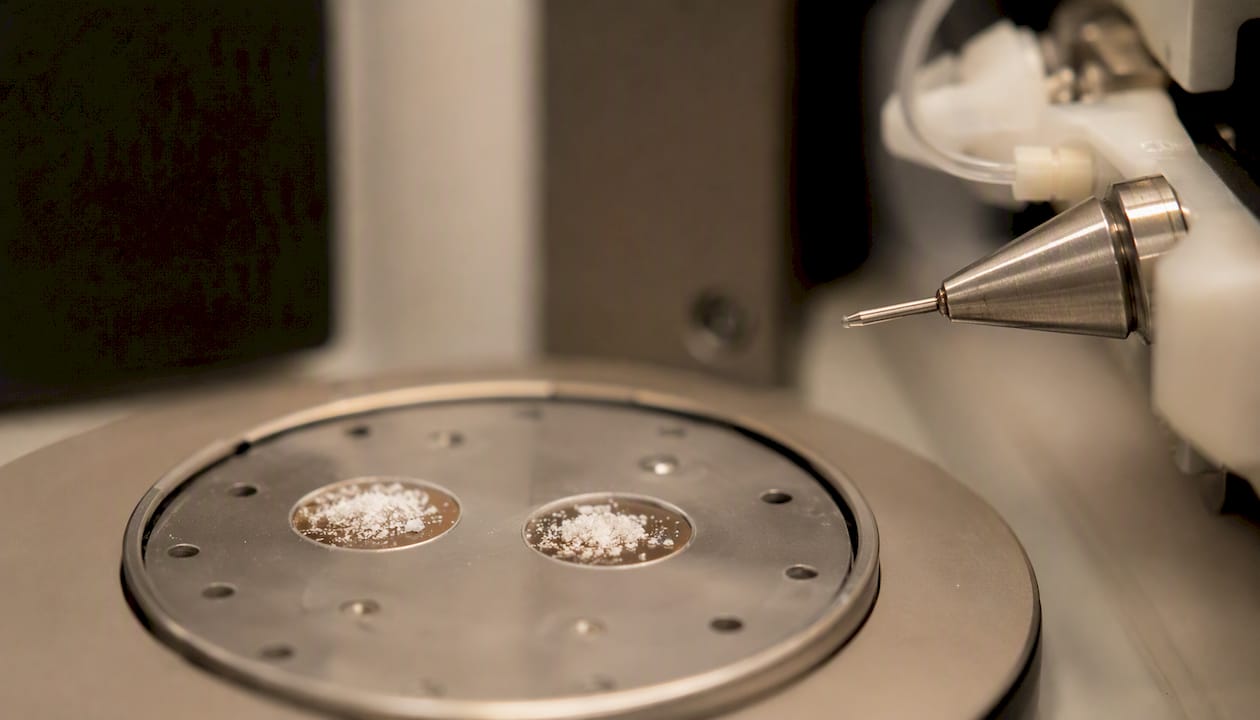
MALDI (matrix-assisted laser desorption/ionization) takes a completely different route. You mix the analyte with a light-absorbing matrix compound, dry it onto a plate, then fire a pulsed laser at the spot. The matrix absorbs the energy, sublimates, and carries the analyte into the gas phase largely as singly charged ions. No solvent flow, no online separation, just a dried spot and a laser pulse.
The mechanism, step by step:
- ESI: charged droplet forms at the capillary tip → solvent evaporates and droplet shrinks → Coulombic repulsion fissions the droplet repeatedly → ion evaporation or charge residue mechanism releases bare, often multiply charged ions into the gas phase.
- MALDI: matrix and analyte co-crystallize on the plate → laser pulse excites the matrix → rapid photoexcitation causes localized desorption → proton transfer in the expanding plume ionizes analyte molecules, typically to a single charge.
That charge-state divergence isn’t a footnote. It’s the single biggest practical difference between the two methods, and it drives almost everything downstream. Multiply charged ESI ions fragment more informatively under collision-induced dissociation, which is why LC-ESI-MS dominates de novo sequencing and PTM mapping. Singly charged MALDI ions, by contrast, make mass assignment simpler because there’s no charge-state deconvolution step, which is exactly why MALDI-TOF remains the fastest route to a molecular weight.
Solvent and matrix choice matter differently for each. ESI performance depends heavily on mobile-phase composition, pH, and flow rate, since those variables control droplet formation and desolvation efficiency. MALDI performance depends on matrix selection (alpha-cyano-4-hydroxycinnamic acid and sinapinic acid are common choices for peptides and proteins, respectively) and laser fluence, since both govern how cleanly the analyte co-crystallizes and desorbs.
APCI (atmospheric pressure chemical ionization) belongs in this conversation too, though it plays a narrower role. It ionizes analytes in the gas phase using a corona discharge rather than droplet formation, and it tends to suit smaller, less polar molecules that ESI handles poorly. For peptide and protein work, APCI rarely enters the picture, but it’s worth knowing it exists as a third option when ESI underperforms on small-molecule LC-MS runs.
A useful mental picture: sketch two panels side by side. Left panel, a charged droplet shrinking and fissioning into a cloud of multiply charged ions streaming toward the LC-MS inlet. Right panel, a dried matrix spot taking a laser pulse and releasing a plume of mostly singly charged ions straight up toward a TOF analyzer. That image explains most of what follows in this guide.
How Do ESI and MALDI Compare Across Key Performance Metrics?
| Dimension | ESI (LC-ESI-MS) | MALDI / MALDI-TOF |
|---|---|---|
| Best for / recommended analytes | Peptides, proteins, complex mixtures, polar small molecules | Intact proteins, rapid peptide fingerprinting, polymers, tissue sections |
| Typical charge states | Multiply charged (2+ to 20+ for large biomolecules) | Predominantly singly charged |
| Coupling to separation | Online LC, direct interface | Off-line spotting after fraction collection |
| Typical mass range / largest analytes | Effectively unlimited via multiple charging; routine up to hundreds of kDa on HRMS | Very high mass tolerance per charge state; intact proteins into the hundreds of kDa range |
| Sensitivity / LOD | Strong with SPE cleanup; sensitive to ion suppression | Good for concentrated spots; less sensitive to trace-level targets |
| Quantitation robustness | Well established, MRM and full-scan HRMS pipelines validated | Improving, but spot-to-spot variability limits routine quantitation |
| Fragmentation / MS/MS friendliness | Excellent, multiply charged precursors fragment richly | Limited without post-source decay or specialized MS/MS setups |
| Sample prep complexity / matrix effects | Requires desalting; prone to ion suppression from co-eluting compounds | Tolerates salts and crude buffers better; matrix crystallization affects reproducibility |
| Throughput / imaging capability | Moderate throughput, limited by LC run time | High throughput per plate; native imaging capability |
Three rows deserve extra attention if you work primarily with peptides and proteins. Charge state governs how much structural information you get out of MS/MS, so if sequencing or PTM localization is the goal, that row alone should steer you toward ESI. Coupling to separation determines whether you can resolve a complex digest at all before it hits the detector, which is where LC-ESI-MS earns its reputation for handling messy biological samples. And quantitation robustness reflects a real, ongoing gap: MALDI has closed some distance here, but validated MRM workflows on triple quadrupoles still set the bar for regulated quantitative work.
Which Ion Source Performs Best for Your Analyte Class?
Performance doesn’t stay constant across sample types. The same instrument that excels on tryptic peptides can struggle with intact glycoproteins or small polar metabolites, and the ion source is usually the reason.
Peptides and proteins. This is ESI’s strongest ground when paired with LC, because multiply charged precursors give collision-induced dissociation something rich to work with, and online separation resolves complex digests before they ever reach the source. MALDI still earns its place here for rapid fingerprinting, intact mass checks on large proteins, and any plate-based workflow where speed beats depth. The split between the two isn’t arbitrary. Ionization efficiency in ESI depends heavily on analyte hydrophobicity, while MALDI ion yield tracks more closely with gas-phase basicity, so the two methods are, in a real physicochemical sense, biased toward detecting different peptides from the same digest.
Small molecules. MALDI can struggle here. Matrix-related background ions cluster in the low-mass region, which can mask or suppress small-molecule signals, and adduct formation complicates spectral interpretation. ESI, often coupled to LC, tends to offer better coverage for polar small molecules, and APCI fills in where ESI underperforms on less polar analytes. Ion suppression from co-eluting matrix components remains a real concern either way, which is why sample cleanup still matters regardless of ion source.
Lipids. Both methods work, but they solve different problems. ESI, run in positive or negative mode depending on lipid class, dominates LC-based lipidomics because it resolves complex lipid mixtures against a chromatographic time axis. MALDI’s advantage shows up when the question isn’t “what lipids are present” but “where are they located,” since direct tissue spotting supports spatial profiling that LC-based methods simply can’t replicate.
Polymers and large biomolecules. MALDI often simplifies interpretation for very large, homogeneous analytes because singly charged ions avoid the charge-state deconvolution that multiply charged ESI spectra require. ESI’s multiple charging, however, is exactly what makes ultra-large species tractable on high-resolution instruments, since it brings enormous molecules down into an m/z range the analyzer can actually resolve.
Peer-reviewed proteomics comparisons consistently report complementary rather than redundant peptide sets between the two ionization techniques, tracing the difference back to amino acid composition, charge-related parameters, and hydrophobicity. Combining both sources measurably increases proteome coverage compared to running either one alone, according to research on ion-source impact on peptide identification.
A separate direct comparison using FTMS instrumentation on gangliosides found that ESI tended to be gentler, preserving more intact ions, while MALDI produced greater fragmentation and different metal-adduct behavior, with negative ion mode performing better for that particular lipid class. The lesson generalizes: ion source choice interacts with analyte chemistry in ways that a single default setting can’t capture.
How Does the Ion Source Interact With Your Mass Analyzer?
Ion source and mass analyzer aren’t independent choices. They shape each other’s performance in ways that catch a lot of researchers off guard the first time they switch platforms.
LC-ESI-MS pairs naturally with QTOF and Orbitrap analyzers in proteomics and pharmaceutical applications, since both instrument classes need a continuous stream of ions timed against chromatographic elution. Where labs do want MALDI-style plate collection from an LC run, they typically split flow post-column and spot fractions manually rather than trying to force MALDI online, since the two ionization physics don’t mix well in a single interface.
MALDI-TOF remains the dominant pairing for MALDI, and for good reason: TOF analyzers measure ions in discrete packets, which matches how MALDI generates ions in laser-pulse bursts rather than a continuous stream. Some hybrid QTOF platforms accept both ESI and MALDI sources on interchangeable inlets, letting a single lab run either mode without buying two separate mass spectrometers, though switching sources still means re-tuning and re-calibrating.
High-resolution analyzers change the calculus further. Orbitrap and FTICR instruments trap ions before measurement, which affects how internal energy from either ionization method translates into fragmentation behavior and how many MS/MS events you can squeeze into a chromatographic peak’s duty cycle. That trapping behavior explains a pattern that surprises newer analysts: even with HRMS instruments now standard in most labs, many pharmaceutical and clinical groups still default to LC-ESI-MS workflows for quantitation, simply because the validated pipeline track record outweighs any theoretical advantage of switching sources.
- MALDI imaging deserves its own callout because it is not a variant of standard MALDI workflows, but a distinct application. Spatial mapping of analyte distribution across a tissue section requires MALDI-TOF or MALDI-Orbitrap configurations built specifically for raster-scanning a surface, and it has no real ESI equivalent.
- Duty cycle and scan speed limit how many precursors an LC-ESI-MS run can fragment before a peak elutes, which is one reason targeted quantitation methods often use scheduled MRM rather than full untargeted scanning.
- Ion trapping behavior on Orbitrap and FTICR instruments affects how cleanly fragmentation spectra come out for either ion source, particularly for labile post-translational modifications.
What Should You Expect From Quantitation and Reproducibility?
LC-ESI-MS quantitation is the more mature discipline, and it shows in the tooling. MRM on triple quadrupole instruments remains the gold standard for targeted quantitation, while full-scan HRMS workflows on QTOF and Orbitrap platforms now provide strong screening capability alongside decent quantitative performance. MALDI quantitation has improved, but spot-to-spot variability and matrix crystallization inconsistency still hold it back from routine use in regulated quantitative pipelines.
Reproducibility problems in each method trace back to different root causes. ESI suffers from ion suppression when co-eluting compounds compete for charge during droplet formation, which is why desalting and SPE cleanup show up so often in method development. MALDI suffers from matrix crystallization differences between spots, since the same analyte concentration can produce wildly different signal intensity depending on how evenly the matrix dried.
Mitigation looks different for each. For ESI, that means solid-phase extraction or other desalting steps before injection, tighter control of mobile-phase gradients, and monitoring for suppression using spiked internal standards. For MALDI, that means standardizing matrix deposition technique (automated spotters help), using internal standards co-spotted with the sample, and averaging across multiple laser shots per spot rather than trusting a single acquisition.
Pro Tip: Stable isotope-labeled internal standards solve a surprising amount of the reproducibility problem for both methods, but the failure mode differs by source. For ESI, spike them in before any cleanup step so they track suppression from sample prep onward. For MALDI, co-spot them with the analyte on the same plate position, since matrix variability between spots will otherwise swamp the correction.
How Do You Choose Between ESI and MALDI for Your Experiment?
Work through these questions in order, and the right instrument choice usually becomes obvious by step three or four.
- Define the experimental goal first. Are you after a quick qualitative ID, deep sequence-level characterization, defensible quantitation, or spatial imaging? Each answer points to a different method before you’ve even looked at the sample.
- Check the sample state. A liquid digest ready for chromatographic separation favors LC-ESI-MS. A tissue section, a crude plate of colonies, or a large batch of similar samples favors MALDI.
- Assess throughput requirements. If you need hundreds of spots screened in an afternoon, MALDI’s plate format wins on raw speed. If you need deep characterization of a handful of samples, LC-ESI-MS’s separation power matters more than speed.
- Evaluate salt content and matrix complexity. Heavy salt contamination that resists cleanup pushes you toward MALDI, which tolerates crude buffers better than ESI does. A complex biological matrix that needs chromatographic resolution pushes you toward ESI.
- Confirm whether chromatographic separation is actually necessary. If your mixture is simple enough to spot directly, MALDI skips a step entirely. If components co-elute or overlap without separation, you need the LC step ESI provides.
- Inventory your instrument access and technician expertise. An LC-ESI-MS run demands column maintenance and gradient optimization skills; MALDI demands matrix selection and plate-spotting technique. Choose based on what your team already does well, when the science allows either.
- Watch for red flags that should flip your default. Need MS/MS-rich, multiply charged precursors and online separation? That’s an ESI signal even if you’d planned to use MALDI. Heavy salt matrix with no time for desalting? That’s a MALDI signal even if ESI was your default.
- Run a pilot on a split sample when the choice is genuinely unclear. Divide the sample, run half through each ion source, and compare identification counts and spectral quality before committing the full study to one method.
When Should You Run Both MALDI and ESI on the Same Sample?
The complementarity evidence isn’t a minor technical footnote. It’s a genuine argument for doubling your analytical workload when proteome coverage matters more than turnaround time. The same 39% overlap statistic cited earlier means that roughly six in ten peptides detected show up in only one of the two techniques, not both.
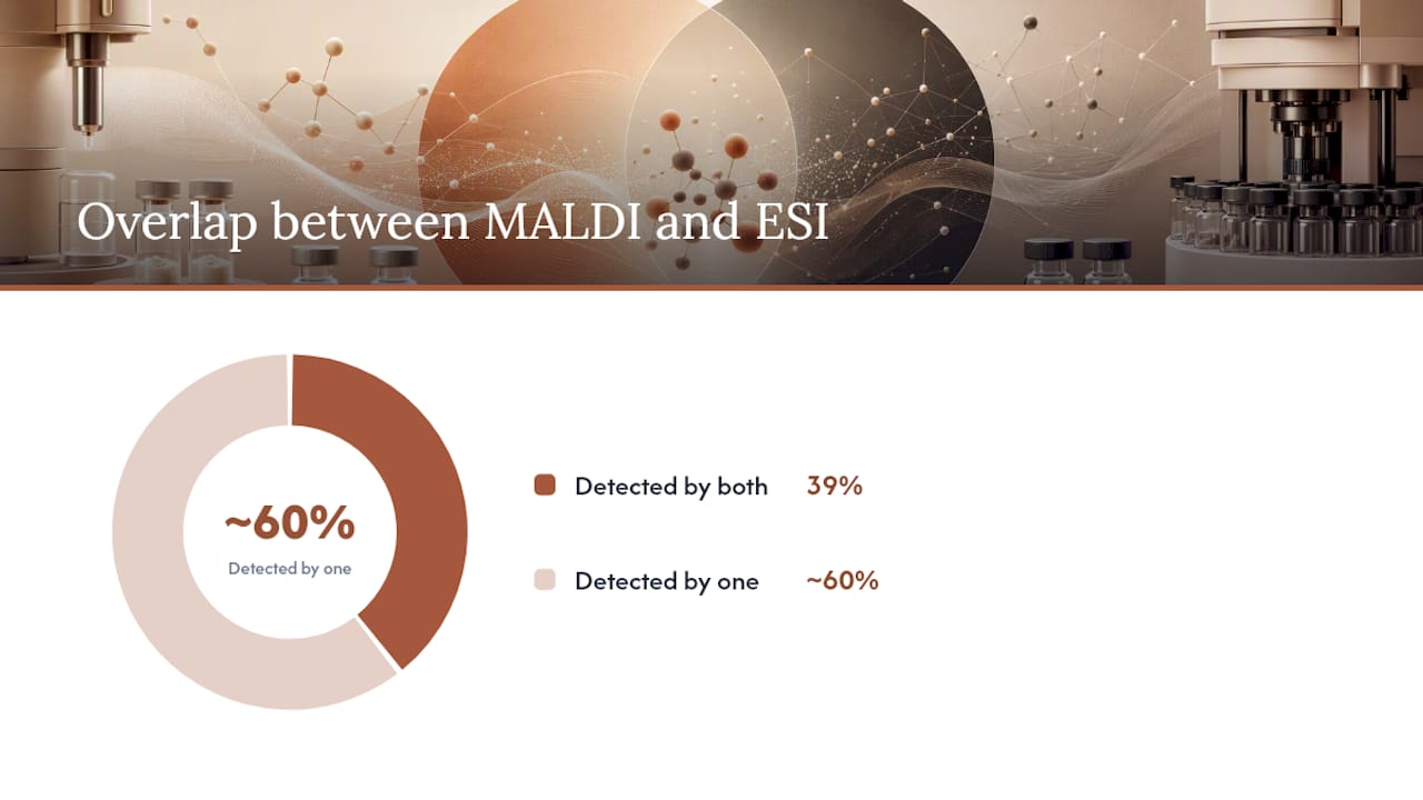
That gap exists because ESI and MALDI respond to different physicochemical properties. ESI ionization efficiency tracks with hydrophobicity, MALDI ionization tracks with gas-phase basicity, and few peptides score high on both scales simultaneously. Running only one method means systematically missing a real fraction of your proteome, not just losing a few edge cases.
Practical ways to combine them:
- Split LC fractions so a portion goes to a MALDI plate for offline spotting while the remainder runs through LC-ESI-MS online, capturing both detection profiles from a single prep.
- Use MALDI first as a rapid screening pass to flag which fractions or samples deserve deeper LC-ESI-MS sequencing, saving instrument time on the slower method.
- Run orthogonal MALDI and ESI analyses specifically for PTM detection, since modification sites often show source-dependent detection bias just like unmodified peptides do.
- Where instrument budget allows, use a hybrid QTOF platform capable of accepting both source types, simplifying data integration since spectra come off comparable analyzer hardware.
Comparisons using a shared hybrid Q-TOF instrument show that ESI interfaces easily with LC and yields multiply charged precursors, while MALDI on the same platform yields mainly singly charged precursors and can suffer suppression effects in genuinely complex mixtures. Running both sources on comparable hardware makes the resulting datasets easier to reconcile than mixing entirely separate instrument platforms would.
Plan for the data integration step before you start collecting spectra. Deciding in advance how you’ll reconcile peptide lists, resolve conflicting charge-state assignments, and merge identification confidence scores across two ionization methods saves considerable rework later.
How Does Ionization Method Choice Fit Into Peptide QC Workflows?
Ion source selection isn’t just an academic question for labs running identity and purity verification on research peptides. It shapes what a Certificate of Analysis actually demonstrates.
That MS step, and MS/MS where structural confirmation is warranted, is where ion source choice becomes a practical decision rather than a theoretical one. LC-ESI-MS delivers detailed identity traces and, where needed, sequence-level confirmation through multiply charged precursor fragmentation. MALDI-TOF-based mass confirmation offers a faster route to an intact mass check, useful for rapid lot screening before deeper characterization.
Each verified lot ships with its own lot-specific Certificate of Analysis, documenting the purity and identity data specific to that production batch rather than a generic specification sheet. That lot traceability matters because peptide synthesis runs can vary batch to batch, and a COA tied to the exact lot in hand tells a researcher what was actually measured on that material, not what was measured on a previous run.
Pro Tip: When evaluating which analytical readout to prioritize for a QC decision, use LC-ESI-MS data when you need purity and sequence-level confirmation with fragmentation evidence, and lean on MALDI-TOF mass confirmation when you need a fast, high-confidence intact mass check across multiple lots. The two readouts answer different questions, and a thorough COA workflow often draws on both.
Researchers evaluating peptide suppliers for laboratory work can review Agateresearch’s research peptide catalog and COA documentation to see how lot-specific verification data is structured and presented for in-vitro research use.
Recommended Reading on ESI vs MALDI Ionization
- Large-scale proteomic study showing limited overlap between MALDI and ESI peptide IDs: the primary evidence behind the 39% complementarity figure cited throughout this guide.
- MALDI versus ESI: The impact of the ion source on peptide identification: explains how amino acid composition and hydrophobicity drive complementary peptide detection.
- Ion formation differences and charge-state behavior for ESI versus MALDI: the mechanistic reference for why ESI produces multiply charged ions and MALDI produces singly charged ions.
- Physico-chemical determinants of ESI and MALDI ion yields: details how hydrophobicity and gas-phase basicity govern ionization efficiency for each method.
- Direct comparison of MALDI and ESI for gangliosides by FTMS: a head-to-head instrumentation study on fragmentation and adduct behavior.
- Ionization modes: ESI, MALDI and coupling to separations: a practical instrumentation review on matching ion source to separation strategy.
- Orbitrap and high-resolution analyzers: implications for ion-source pairing: covers how trapping analyzers interact with ion source choice for MSn workflows.
- Comparison of ESI and MALDI on the same hybrid Q-TOF: implications for complex mixtures: a same-instrument comparison showing suppression effects and precursor charge behavior.
Sources
- Large-scale proteomic study showing limited overlap between MALDI and ESI peptide IDs
- Ion formation differences and charge-state behavior for ESI versus MALDI
- MALDI versus ESI: The impact of the ion source on peptide identification
- Ionization modes: ESI, MALDI and coupling to separations